Alena Pance from our Public Engagement team introduces the Human Cell Atlas and a new animation explaining the SARS-CoV-2 infection process.
The quest to identify and characterise every cell in the human body sounded like science fiction a decade ago, but with fast advancing technologies more and more scientists all over the world are joining in this venture. The Human Cell Atlas consortium (HCA) has as primary goal to create a cell map of the human body that will provide fundamental knowledge to understand how it works. This will lead to a hugely detailed picture of what normal, healthy cells and tissues are like, which will form the basis to help identify, monitor, and ultimately treat disease.
The Wellcome Genome Campus contributes strongly to the Human Cell Atlas effort with the participation of several groups from the Wellcome Sanger Institute as well as the EMBL-EBI. In the Sanger Institute, many of the participating groups are in the Cellular Genetics Programme, led by Sarah Teichmann who is also the co-chair of the Organising Committee of the HCA.
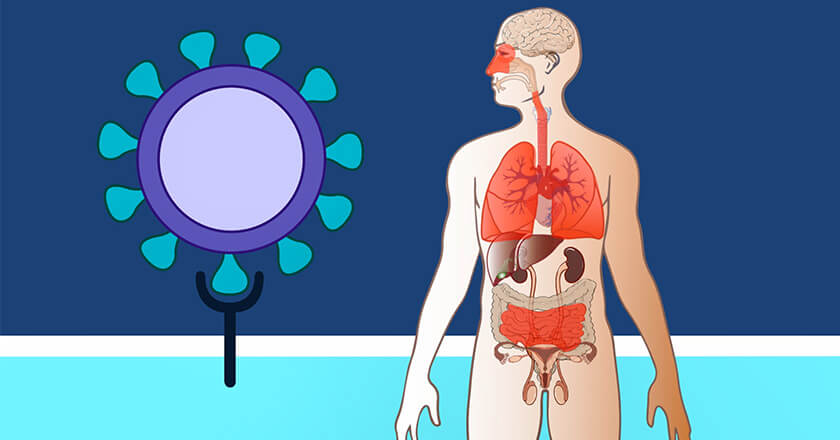
In a rapid response to the pressing public health situation at the beginning of the year, biological data produced by the HCA was mined to investigate the expression patterns of genes (which genes are turned on or off in a cell, and when) in human cells infected by the SARS-CoV-2 virus. A particular focus of the study were the genes that make two key proteins, ACE2 and TMPRSS2, which are involved in the entry of the virus into human cells. Given the importance of the airways in the development of the disease, the work initially concentrated on the lung, but quickly evolved to broaden its scope. Interestingly, the expression of both these genes was detected in many cell types, including nasal and corneal cells, and those lining the intestine and nose. This pattern of expression may help to explain the route of Covid-19 infection, transmission and spread.
From its foundation the HCA has been committed to sharing its results and discoveries as widely as possible. As part of this mission, the Wellcome Genome Campus Public Engagement team have been supporting the HCA in their applications for grants specifically funding public engagement and citizen science.
One of these successful engagement grants focuses on fostering conversations between HCA scientists and artists about HCA research, to stimulate discussions and interpretations of emerging issues including health, ethics, science and more. As part of this work, Jana Eliasova, Human Cell Atlas medical illustrator, and Nick Lewis, Lecturer in Illustration, Animation and Games Art at Sunderland University teamed up with Suzy O’Hara, project manager for the art-science collaboration and Paul Gibson, Project Manager for Cell Atlasing, to create an animation for young people (Key Stage 3), explaining how HCA scientists are helping the global fight against the COVID-19 virus.
This video is the first of many anticipated outcomes of this scientist-artist collaboration.
For more information, and opportunities to get involved, please visit humancellatlas.org/wellcome-engagement .